Your dog suddenly stops eating, or your cat begins hiding and showing signs of discomfort. These changes can be concerning, especially when the cause is not immediately clear. Some health problems develop inside the body without producing visible symptoms.
For pet owners in Weaverville, understanding veterinary imaging can help them make informed decisions about their pets’ health. At Broad River Animal Hospital, diagnostic care for pet health includes digital X-rays, ultrasound imaging, and laboratory testing to investigate medical concerns.
X-rays and ultrasounds help veterinarians diagnose pet health issues by revealing internal structures that a physical examination can’t fully evaluate. These imaging methods provide different information about bones, organs, and surrounding tissues, helping veterinarians determine appropriate next steps.
How Do Veterinary X-Rays Detect Health Problems in Dogs and Cats?
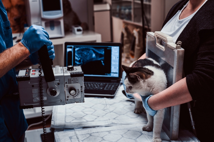
Veterinary X-rays, also called radiographs, use controlled amounts of radiation to create images of structures inside an animal’s body.
They are particularly useful for examining bones, the chest, and certain abdominal conditions.
Veterinarians may recommend X-rays to investigate:
- Bone fractures: Identifying broken bones and assessing their alignment.
- Joint abnormalities: Detecting changes associated with arthritis or injuries.
- Breathing difficulties: Examining the lungs and surrounding chest structures.
- Heart enlargement: Evaluating changes in the heart’s size and shape.
- Swallowed objects: Identifying certain foreign materials or signs of intestinal obstruction.
- Abdominal abnormalities: Assessing organ size, abnormal gas patterns, or possible masses.
Digital radiography produces images quickly, allowing veterinarians to examine areas of concern without waiting for traditional film processing.
However, X-rays cannot identify every disease. Additional testing may be necessary when abnormalities involve soft tissues or internal organ function.
What Can an Ultrasound Reveal About Your Pet’s Internal Organs?
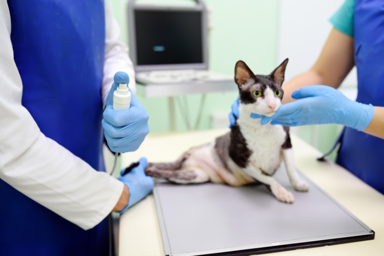
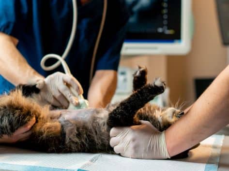
Ultrasound imaging uses high-frequency sound waves to create moving images of internal structures. Unlike X-rays, ultrasound does not use ionizing radiation.
It allows veterinarians to examine the appearance, structure, and movement of certain organs in real time.
Ultrasound examinations may help identify:
- Changes in the liver, kidneys, spleen, or bladder.
- Abnormal fluid accumulation within the abdomen.
- Certain tumors, cysts, or abnormal tissue growths.
- Changes in intestinal walls or other digestive structures.
- Urinary tract abnormalities, including some bladder stones.
- Pregnancy and fetal development.
Ultrasound can also help guide certain diagnostic procedures when additional tissue or fluid samples are needed.
Because sound waves do not travel effectively through gas or bone, ultrasound has limitations when examining air-filled lungs and structures hidden behind bone.
X-Rays vs. Ultrasounds: Which Imaging Test Does Your Pet Need?
Neither imaging method replaces the other. Each provides different information depending on the suspected medical condition.
| Veterinary X-Rays | Veterinary Ultrasounds |
| Particularly useful for bones and chest structures | Particularly useful for abdominal organs and soft tissues |
| Produce still images | Produce moving, real-time images |
| Use a small amount of ionizing radiation | Use sound waves without ionizing radiation |
| Help identify fractures and certain obstructions | Help evaluate organ structure, fluid, and certain masses |
For example, a dog with suspected intestinal obstruction may initially need abdominal X-rays. Ultrasound may then provide additional information about the intestinal walls or surrounding tissues.
Sometimes, veterinarians recommend both examinations to investigate a medical concern more thoroughly.
When Should Your Pet Have an X-Ray or Ultrasound?
Not every unusual symptom requires imaging. Veterinarians consider the pet’s medical history, physical examination, and clinical findings before recommending diagnostic tests.
Schedule a veterinary examination if your dog or cat develops:
- Repeated vomiting or diarrhea.
- Persistent loss of appetite.
- Unexplained weight loss.
- Limping or difficulty moving.
- Abdominal swelling or discomfort.
- Changes in urination.
A single mild symptom in an otherwise active pet may sometimes be monitored with veterinary guidance. Persistent or worsening symptoms require evaluation.
Difficulty breathing, collapse, severe abdominal swelling, repeated unproductive retching, or inability to urinate require immediate veterinary attention. Do not delay emergency care while waiting for a routine imaging appointment.
What Happens During a Veterinary Imaging Appointment?
The examination generally begins with a review of symptoms and a physical assessment.
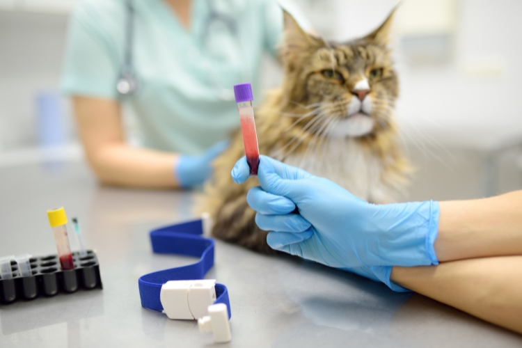
Dr. Allison may consider imaging findings alongside bloodwork, urinalysis, or other diagnostic information when evaluating a pet’s condition.
During X-rays, pets must remain positioned correctly for clear images. Ultrasound typically involves applying gel to the skin and moving a handheld probe across the examination area.
Some pets require sedation to remain comfortable and still. Others can undergo imaging while awake.
Call our animal hospital now if your pet has concerning symptoms and you are unsure whether diagnostic imaging is necessary.
How Does Diagnostic Imaging Help Veterinarians Plan Treatment?
Imaging findings can help veterinarians determine whether a pet needs medication, additional testing, monitoring, or surgery.
For example, identifying a fracture may guide orthopedic treatment, while detecting abnormal abdominal fluid may prompt further investigation.
Imaging alone does not always establish a final diagnosis. Combining diagnostic care with laboratory results and physical findings supports more informed medical decisions.
Get Answers About Your Pet’s Health With Veterinary Diagnostic Care
Unexplained symptoms deserve careful attention. Diagnostic imaging helps veterinarians investigate internal health concerns and identify changes that may not be apparent during a physical examination.
At Broad River Animal Hospital, we provide diagnostic care for pet health in Weaverville, NC, including digital radiography, ultrasound, and in-house laboratory services.
If your dog or cat is showing unusual symptoms, call our hospital at 828-484-7771 to schedule a veterinary examination and discuss the appropriate diagnostic tests.
Frequently Asked Questions
1. Are X-Rays Safe for Dogs and Cats?
Veterinary X-rays generally involve brief, controlled radiation exposure. Veterinarians consider the clinical need before recommending them. Proper positioning and imaging techniques help minimize unnecessary exposure.
2. Does My Pet Need Sedation for an Ultrasound?
Many pets undergo ultrasound examinations without sedation. However, anxious, painful, or restless animals may need additional support. Our doctor at Broad River Animal Hospital determines whether sedation is appropriate based on the pet’s condition.
3. Can an Ultrasound Detect Cancer in Pets?
Ultrasound may reveal abnormal masses or changes in internal organs, but it cannot always determine whether a growth is cancerous. Additional tests, such as tissue sampling, may be necessary to establish a diagnosis.
4. How Long Does a Veterinary X-Ray Appointment Take?
The imaging procedure itself is usually brief. However, positioning, preparation, sedation when necessary, and image interpretation can extend the appointment. Our veterinary team can explain what to expect.
5. Can Blood Tests Replace X-Rays or Ultrasounds?
Blood tests assess certain aspects of organ function and overall health, while imaging examines physical structures. These tests often complement each other rather than replace one another.
6. Should Senior Pets Receive Diagnostic Imaging Regularly?
Senior pets may develop conditions that require imaging, but routine X-rays or ultrasounds are not necessary for every animal. Our team at Broad River Animal Hospital recommends tests based on examination findings, medical history, and individual health risks.